Exploring Visceral Surfaces
Description
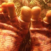
Gil compares the dissection of an unfixed form to a fixed form while looking at the viscera.
This video was filmed and produced by Gil Hedley. Please note that it includes graphic videos and photos of dissections of cadavers (embalmed human donors). You can visit his website for more information about his workshops.
About This Video
Transcript
Read Full Transcript
Hi, my name is Gil Hedley, and thank you for your interest in the Integral Anatomy Series. This fourth volume of the series is devoted to the viscera, their relationships, and their continuities. The skin, superficial fascia, deep fascia, muscle, bone, and serous membranes are the anatomical context for understanding the viscera. The initial volumes of the series provide that context. Watch them first if you are committed to understanding the viscera, as they provide the essential framework for comprehending what follows.
When I enter the lab, I do so with appreciation for the gift which I am receiving. As we explore in the lab together, I invite you to share in my gratitude for the donors and their families, as we who view this material are the direct beneficiaries of their generosity. I place no particular premium on fresh tissue dissection over dissection of embalmed tissue. There are advantages and disadvantages to both, and neither can lay claim to representing perfectly the living miracle of the human form. Both are methods which grant us different kinds of access and insights.
Embalming allows a slow and methodical dissection and enhances our ability to see certain tissues like the deep fascia more clearly, and embalming nicely fixes the shape of the viscera for our observation, though the colors and the textures it generates are somewhat off. Fresh tissue offers some insights into the textures and colors of the body which are lost to the preservation process, but it offers a much shorter time frame for observation, and it can be even more difficult to present with respect to the aesthetics of the images created. With these considerations in mind, I invite you to observe with me this unpreserved form of an elderly woman. The onion-tree model is a functional simplification of the human body. With it, I can reference the whole mass of the viscera considered together as a deep layer of the onion, or as the branching trees as well, at least in the case of the neurovascular trunks and limbs.
The skin itself is the terminus of those visceral branches from the neurovascular trunks as they interface directly with the external environment of the body. The superficial fascia is a great suspensory web of perception of a particular frequency range within which the neurovascular pathways braid their way out from the core and envelop our whole being amidst the yellow finery of our sensory fleece. We can separate out tissues and layers and pathways of connection which we hold dear to our mental conception of the body, but the models conceal the whole as much as they point to it, to the extent that we sometimes come to prefer our models to the reality at hand. The fact of the whole remains in spite of our attempts to parse the whole body into tissues or layers. The viscera are not limited in their physiological function or in their anatomically demonstrable extent to the thorax or the abdomen or the cranium, though through the conventions of our training we may habitually limit our ability to see them beyond their designated regions.
From an integral viewpoint, the viscera are considered to be non-local phenomenon, extending in both form and function through all the textural layers of the body. They are commingled with all the tissues of the body, so that with all fairness we can speak of the viscera of the arm, or the viscera of the leg, or even the viscera of the fascia, surely to the extent that these tissues are innervated and vascular. The motions of peristalsis and the rhythmic pulsations of the heart and the cascading waves of the brain all reiterate and reverberate and enliven the whole body and are in mutual relationship with the forms and processes of the other layers. If a massage hydrates tissue, increases circulation, and induces relaxation, it is in some direct measure because the viscera have been touched, right there in the muscle tissue and fascia of the arm or the leg or the back or the belly. Every massage therapist and body worker is doing a form of visceral work.
That pulse in the wrist is the arm of the heart. When you stroke the skin, you are literally caressing someone's brain. Touch is an intimate responsibility for the professional because it is only possible to touch the whole person. So we're working our way down through the superficial fascia of this form and encounter a texture that's quite different from the surrounding superficial fascia. We see that it's at the base of an old incision and what we're looking at is the herniation of some intraperitoneal fat bulging through the base of that incision still covered in peritoneum.
As we inspect it, we can notice its smooth and shiny texture is quite different than the more bubble wrap-like consistency of the surrounding superficial fascia. The inner form of the deep fascia woman here is seen emerging from the adiposal organ at this point only partially dissected. The presenting morphology is quickly transforming. The superficial fascia is a shaping layer, as are the muscle, viscera, and bony layers. When we draw it away from the body, an utterly distinct morphology presents itself to view.
Dissected away from the body completely and viewed independently, the common texture and substance and mass of the superficial fascia is here appreciated as an anatomically and physiologically distinct organ in its own right. I wasn't sure when I initiated this dissection whether or not the unpreserved tissue would lend itself to this approach. In fact, it was no more or less challenging to accomplish. The integrity of the superficial fascia is what creates the possibility of this dissection. Pressed by the contingencies of the unpreserved tissue, a patchwork of superficial fascia or remnants overlays the deep fascia, which is nearly translucent in the upper body especially.
In the lower limb, the deep fascia is a more thickly woven web of fibers. Looked upon closely, the minute branchings of the heart are present. At no point from the center of the chest to these tendrils feeding the deep fascia of the leg, do we find any interruption of the flowing continuity of the viscera as they perfuse every textural layer of the human form. When you were first introduced to the form of this elderly woman a few minutes ago, you might not have imagined that this would have been her shape at the level of deep fascia and muscle, the primary presenting layer which shaped her form was her superficial fascia. If you were an artist drawing the lines of this body, it would be the shape of the superficial fascia which you would need to account for to render her accurately more so than the muscle.
Though I have been saying that the viscera reach beyond their spaces into the layers surrounding them, the organs in this particular body do not define the woman's shape as they sometimes do. Learning to tell which layer is primarily shaping someone's body is a practical undertaking. The shaping layer may offer clues about a given person's life experience. When observing a person's body, it's a fair question to ask then what layer is presenting itself to my attention when I observe this person's shape. If it is the superficial fascia, you would probably do well to attend to its particular needs as opposed to skipping past that layer because your personal or professional experience may be to work with the muscles or the organs or the fascia construed as distinct objects of touch.
When each textural layer is given its due, the possibility for an integrated experience of one's body is built into the approach. Having noted the new form is primarily shaped by the superficial fascia, we reintroduce the male form with which we are already familiar, noting that his body primarily presents the shape of his muscle layer defined by his deep fascia. With the superficial fascia removed from them both, the unemboned form and the male example look surprisingly similar. Both have concave abdomens. Neither form present the viscera prominently, as does the female form from the first volume, whose viscera present clearly in her gross morphology.
Her abdomen is more convex than concave with or without the superficial fascia. So among these three examples, we see one shaped particularly by the superficial fascia, one by the muscle, and one by the viscera. When attempting to palpate the viscera of people with different gross morphologies, it is important to note the variations which create the anatomical context for such work. When leveraging the bony layer to create tension in the deep fascia of the abdomen, we can create vectors of pull generated by torsion and watch the translation of the movement through the scars in the anterior rectus sheath. We're not often privy to witnessing these normally hidden motions.
So I'm incising the tendon of the external oblique somewhere along the inguinal line, I like to call it. So as I reflect through on back to the anterior superior iliac spine, I reveal that deep to the external oblique fascia here, we immediately see the muscle tissue of the internal oblique. But I'm going to make a big sweeping circle actually, to increase our window size. So in a sense, we can also say I've got the anterior erectus sheath in my hemostat right now. And the tissue is, although not embalmed, if I'm careful, don't hit any major vessels it will stay pretty clean for our view.
But now I'm coming into scar tissue here, you see how it doesn't want to yield to my scalpel anymore? How smooth and easy the tissue is differentiated here, and then I come up to there and all of a sudden you can hear it scratchy, and there's a network of collagenous fibers that have woven in and tacked the layers down to each other here, and I'll have to scratch my way through them and dull my blade on scar tissue and nylon. So now we've displaced that external oblique and its fascia, we've exposed the rectus abdominis, we can see that this rectus abdominis has been excavated, right? That fatty deposition there really represents the filling in of the rectus abdominis, which was cut. The rectus abdominis looks like it didn't survive the surgery.
You mean that they actually removed some of the muscle fibers, and so fat has now replaced? Exactly, it's filled in the space. So here's rectus abdominis in its fullness, but this one was intruded upon. We have muscle fiber, muscle fiber, muscle fiber, and then a gap. So now I'll lift the remnant of that rectus abdominis, now there's more stitches underneath that we didn't see before, so they'll create stitches here and then displace the muscle tissue, cut through, stitch underneath it, and on their way out.
Well at least they did it in layers. Yeah, that's right, they do. They do it in layers, and then they have a great consciousness, so the surgeons are incredibly attentive to all the layers that they've gone through to make sure that they put them all back together. Dropping rapidly in through the layers already covered in the other DVDs, we see here the beautiful sheen of the glistening parietal peritoneum. As I differentiate that serous membrane from the overlying transversalis fascia to which it is normally adherent, the membrane is thin but resilient and encompasses the majority of the abdominal viscera.
Having brought you into the dissection of the unembound female form to the same layer that we had reached in the embalmed male form, we return our attention now to that male body to continue where we left off and begin our tour of the viscera at their center. We will return our attention to the unembalmed female form again later. Her body is both instructive and riveting, but it is important first to establish an understanding of more normal anatomical relationships as we can in this male example before exploring the artifacts of surgery which influence the relationships of the viscera in the female form. See, we have the stomach and the stomach's skin here, and then as I draw down on the greater omentum, we can see how the greater omentum is coming off of the greater curvature of the stomach here, clear around the bend, and so this is greater omentum also, all around the greater curvature of the stomach. I'm drawing up on the greater omentum the four layers of peritoneal wrap contributed by the transverse colon and the greater curvature of the stomach along the greater curvature of the stomach to create this four-layered intelligent fabric, this internal organic snuggle blanket.
This is a more typical presentation of the greater omentum, contrary to the current wisdom, I do not consider the fatty deposition here to be a de facto indicator of ill health. As a matter for consideration, the greater omentum might be investigated as a sort of internal poultice with absorptive properties which, like a clay poultice, has the capacity to draw off toxins and reduce inflammation in the tissues it contacts as it migrates throughout the abdomen like a mobile lymphoid organ or an itinerant country doctor. I've watched students commenting on the fatty tissues they find inside the abdomen conclude the person must have been unhealthy only to learn the donor died in their late 90s and lived a very active life. The fatty lobes revealed along the transverse colon are deposits within the transverse mesocolon. We saw the same types of deposits in the embalmed female form.
Looking closely, we can see the visceral layer of the peritoneum overlying the transverse colon and extending to form the membranous structure of the greater omentum. At this point, we can see very clearly the cone sweeping down, it bends, it flexes, that's the flexure and where is it flexing, it's flexing at this organ that we saw from behind and there we can see it. Down here it's the spleen, see the spleen, I just popped the peritoneum out from around the spleen hooked in at the flexure to the transverse colon, at the splenic flexure of the colon, transitions from the transverse colon to the descending colon at this point and over here we have our splenogastric ligament, the peritoneum spanning across from the spleen here to the stomach because the stomach is really big, it goes all the way around to the back. We saw this tiny little bubble here, the surface projection of the stomach, this tiny little bubble, there it was and we said that's the stomach and you're like what and here's the gastric impression, the impression that the stomach makes on the liver, hanging out there for 70 years together and it makes an impression on each other. So there's the stomach but that's just a little tiny bit of the stomach and you pull it back apart and you see this whole tremendous fabric here of the stomach and we have our spleen.
Okay we noticed also this ligament running between this spleen and the liver so we have our hepatosplenic ligament and our splenogastric ligament and our splenocolic ligament here. So the spleen is like a slingshot coming through these ligaments coming off of the liver and the stomach and the colon and we're seeing that at this point there's an arc of tissue and this arc of tissue is the hiatus in the diaphragm through which the esophagus passes right. It's a beautiful shot of the hiatus here. So a hiatal hernia is when this tissue, this arc of the diaphragm loses its integrity for some reason and the stomach which is trying to get sucked up into the thorax after all anyway but is usually blocked by the diaphragm. It goes through this space and a bubble of the stomach, usually this upper portion here, a bubble of the stomach will slide up into the thorax through this arc of tissue here, this arc of tissue in the diaphragm.
So at this point the pathway of your food transitions from the thorax to the abdomen. That's neat. We can see the, get a sense of the expanse of the stomach and see that the stomach is also jointed with the spleen. We were marking about the joint of the spleen with the lung but here we can see the interior surface of the spleen is jointed with the stomach. Those are sliding surfaces there.
See the spleen and the stomach. The spleen sliding on the stomach and each breath, the spleen and the stomach are sliding relative to each other. The spleen and the lung are sliding relative to each other. The stomach and the lung, the stomach and the liver, fabulous. So I draw down my stomach here, the fundus of the stomach and I trace along this great bag and then it gets very skinny over here.
The bag gets skinny, the pyloric portion of the stomach and as we get over to here and to the pyloric valve itself. So it's quite an expanse, the stomach has got to be a foot across practically and it's arcing this way around the form and it's also in this plane here. So it's an arced, the greater curvature of the stomach is an arc following the arc basically of the costal margin as well. So we have the, oh it's actually above that, it's higher than that, the stomach is right inside your ribcage just like the liver is on this side. So the stomach of course can drop down, if it's really filled up the whole sack can sort of sag.
You eat Thanksgiving dinner and your stomach bloats and sags and it can be hanging out down over your intestines here. I've seen that in radiographists where the stomach is you know sagging down into the pelvic brim even. At this point we see the peritoneum coming across to the, this left lobe of the liver and it sort of gets into this little point here and that's called the left triangular ligament of the liver and then here we see the continuation of the, here's the parietal or the wall layer of the peritoneus coming here and it's diving down underneath the heart here and it sweeps down to the liver all along this, it crowns the liver, there's a corona across the liver and that we call the coronary ligament of the liver. The falciform ligament I'm cutting at this point sort of like a septum that comes from the parietal peritoneum down to form the skin of the liver and then this cord like terminus of the falciform ligament which actually comes clear underneath here. You can see it's kind of a cord, it's like a cord, it's roundish and it's like a vein but it's not a vein at this point, it was a vein in the fetus but now it's more like a cord, it's the round ligament of the liver and it makes its way onto the navel.
So we have the coronary ligament on the right side here, it's just the peritoneum sliding down to form the skin and the coronary ligament on this side, the peritoneal sac has been drawn away, tucked away here. I can see that the liver is here again having a point where the skin of the liver comes up to form the peritoneal sac. So the parietal peritoneum is transitioning with the visceral peritoneum, the skin of the liver at this point and this would be the right triangular ligament of the liver. If I cut through this point, we'll demonstrate that the peritoneum itself is coming from either side here. You see the peritoneum itself is as it folds around and this little gap here in the covering of the liver, if I push it away, will represent the bare area of the liver where the peritoneum didn't manage to squeeze over and form a skin there.
We have the round ligament, the falciform ligament, the right triangular ligament, the left triangular ligament and the coronary ligament and the actual liver tissue is red and supple and soft. Here it's embalmed and toughened and has coagulated blood in it so it doesn't have the same texture but what a glorious shape it has and I'm really wanting to explore these shapes and the relationships of the organ and all that. The liver is dropping down from the contraction of these diaphragm fibers. The liver is pushing down, displacing the abdominal organs and this margin of the liver will drop past the margin of the ribcage, the ribbasket, the costal margin here. It will drop down past it and the liver will expose itself from behind the bone and because it has this ligament here, the coronary ligament and the triangular ligaments that we've been indicating, the liver is rocking forward along this orientation of its triangular ligaments, and it's doing a motion like this.
Why that motion? Well, because it wants to, I don't know, because the motion is defined by the peritoneal relationships. So why are we bother learning about these ligaments? It gives us a sense of how our organs, their mobility, the way that they move around inside of the visceral space relative to one another and the motility would define the motion of the organ within itself, its own intrinsic life activity. And then if I lift up, now we've explored the top of the liver, here's the bottom of the liver.
Okay, so we see this gallbladder and the gallbladder was tucked here and we kind of flip it out a little bit and we see it's this nice sack. And is it broken? No. No. People think, oh, it's all green in here.
It must be broken. But really, the biliverdin of the bile has sort of perfused this whole form at this point. So things are a little green. You see this little hole here, this little window? I didn't put that there.
That's there normally. But I'm indicating it because I'm pointing out this ligament here, the hepato-duodenal ligament. Okay, and the hepato-duodenal ligament is simply that skin of the liver now coming off of the liver and heading on over to cover the duodenum and join up with the stomach here as well. And if I go through this little foram, this little opening, this little famous opening, this little opening, I'm going beneath something. See there's like a, I'm tugging up now.
There's something to tug here. Okay, there are three structures in there. Well, four. It's the hepato-duodenal ligament, of course. But it's also the pathway from the liver to the duodenum along the peritoneum is called the hepato-duodenal ligament, but the common bile duct is running through here, the common bile duct, the left hepatic duct and the right hepatic duct, the cystic duct.
These are all passageways for bile. The three form together in a common union, the common bile duct, and I can sweep my finger under here and I'm underneath now the bile duct going from the liver and the gallbladder, the common bile duct to the duodenum, which is where the bile empties in. There's two more structures here. The hepatic artery, which is the blood supply to the organ of the liver. And then there's the portal vein.
A portal vein is the drainage of the entire gut here to the liver. So all that stuff you eat is going into the intestines, the nutrition and toxins as well are being drained out through the portal system and they're all converging to the portal vein into the liver right through here. It's hidden behind this ligament, but I haven't dissected anything out yet. Again, we're looking at everything in place first. Then when you take it apart, you know what happened.
So that's what we're doing here now. There's more, because if I look here now, I see I'm seeing more of my stomach in place. I'm lifting up my heart here. I'm pulling back my liver so we can peek into this place and my gosh, look, we see this stomach here, the pylorus is this tough rubbery part here, the pyloric portion of the stomach and then the pylorus itself or the pyloric valve. And then as I draw the stomach down, I see this kind of a, another filmy fascia here, so many filmy fascias in the body, a little filmy fascia.
Now if I get my hand underneath that forearm and that opening, I can see, look, my finger is behind that filmy fascia, a filmy fascia going from the stomach over to the liver. It's spanning between the lesser curvature of the stomach, the small curve of the stomach here, this is the big curve of the stomach, this outer curve, the big curve of the stomach is called the greater curvature and the smaller curve of the stomach on the inside is called the lesser curvature. So this would be called the lesser omentum. It's the peritoneum, the skin of the liver and the skin of the stomach connecting to each other and creating a span, a little circular span of tissue here between the two. So I can sneak into that epiploic foramen of the lesser omental bursa, we're getting really technical here, and I sneak my finger underneath the artery and the vein and the common bile duct and I get underneath and now I'm in like a, I'm in a space, it's the space behind the lesser omentum, it's very thin, you can see the blue of my glove here and actually my finger's on a pancreas now because when you draw the stomach down, when you draw the stomach down and you see this lesser omentum here, this little sac, deep to it is the pancreas.
As a point of comparison, let's study now the liver of the unpreserved form. Now we see much more of the liver, there's a little adhesion, there's some adhesions of the liver, I'm not surprised to see more adhesions and here's the remnant now, that falciform ligament that I indicated a moment ago and there are adhesions, see I'm pulling away these adhesions and even as I do so, I disrupt the liver tissue just a little bit doesn't yield so easily as I pull away the adhesions but this falciform ligament, then this is normal, see we have the adhesion, I pulled that away but now we have a relationship of the liver through here so we have the skin of the liver rising up into the diaphragm covering the peritoneum, the parietal peritoneum here swooping down over the liver to the skin of the liver so as we follow along the crown of the liver, here's another adhesion by the way, see that adhesion, this should be a sliding surface but here the visceral peritoneum covering the liver, the skin of the liver is adherent to the parietal peritoneum, I can break it with my finger and as I do so I get to scoop underneath the mass of the liver and what an enormous and beautiful organ it is and as I displace it laterally I see its connection here to the diaphragm through the peritoneum, the liver and the diaphragm have an intimate relationship, the diaphragm is like a stocking cap over the liver so every contraction of the fibers of the diaphragm represent a motion for the liver and as I slide that stocking cap off of the liver and reveal the organ itself, we see it has more relationships in the back here, along here it's like a tiara or a crown, we call it the coronary ligament and at its lateral extent we call it the right triangular ligament here. This tissue here of course is peritoneum but it's covering an organ, the kidney, the kidney in its subserosal fascial packing has a sliding surface right here with the liver, isn't that beautiful? Look at that supple organ sliding against the kidney and then back up here we have that relationship with the liver to the colon, so here's the liver to the colon and here's the liver to the diaphragm and the triangular ligament on the right side and if I brush away here you'll see more of this massive organ, look at how it extends clear across her body, it's almost to the rib on this side I can feel the tip of the 11th rib with my thumb right here and I see the tip of the left lobe of her liver, now I'm not telling you this liver is enlarged only that it's a big organ, big and soft. Well you know what I'm noticing?
That's a little different here in a normal configuration of a body, is that the greater omentum is adherent to the bottom edge of the liver, isn't that different? Because the greater omentum is an organ that's coming from the stomach and the transverse colon but in her body the greater omentum is adhering to the lower margin of the liver, so if I push some more, look now I'm pushing, I'm pushing, I'm pushing, what am I pushing? I'm pushing the stomach which is also adhered here to the liver, the liver and the stomach normally have a sliding surface but in this form the stomach is adhered to the liver, the greater omentum is adhered to the liver, the greater omentum is adhered to the peritoneum, so we have a lot of adhesions in this form. As I continue to push though I can free the bottom, the gastric impression of the liver from the stomach itself and get a sense of that organ and its difference. So this sac here is being revealed now, you see we have a greater omentum and we have this different color, here we have the yellow and here we have the red of the liver and then there's a different color here and that's the muscle wall of the stomach.
Now we'll return to the preserved male form, the loops of the small intestines are all piled down here, they're numerous, loops and loops, see here we do get the rounder look, scoop the ones out of the deep pelvis here, more loops, thought there was nothing there for a minute huh, but now look at them, more loops, there's no shortage of intestines here huh, oh my goodness, look now I have this tremendous bouquet, where did they come from? Well they were lying in their spaces, the deep pelvis, across the brim, scooped in down here, right down to the large intestine, I'm holding the bouquet of the intestines and I can rock his body with the anchor of the small intestines, which is known as the root of the mesentery, so I've gathered up the loops but I've also gathered up the mesentery, that's the mesentery, the mesentery is the fabric along which this set of loops, rides, the mesentery is this fabric here, now the fabric is made up of the peritoneum, except here it's called the mesentery, so we have a layer in the front, or a layer on one side and a layer on the other side coming off the skin of the organ in the same way that the greater omentum was formed, we have this skin of the organ here, the visceral peritoneum, and then the sheets here of peritoneum that form this fabric, this fabulous fabric here, like a sea plant, this fabric is called the mesentery, the mesentery has its root, meaning it arises from the back of the abdominal space, as it comes up and arises to form this complex sheeting, running through the mesentery are blood vessels, I'm backlighting the mesentery, so you can see how arising from the intestine and through the mesentery are three trees, the lymphatics, the arteries and the veins, there's a nerve tree in there too, so a lot of four trees, I'm just going to keep going, I'm going, I'm going to put the whole tube pass through my hands here, and you'll see that no matter where I go on this fabric, I have the intestine, the elementary canal sort of riding along the top, and its blood supply passing through the mesentery towards the root, now two main arteries feed the mesentery here, the superior mesentery and the inferior mesentery, and one great vein drains it, so all these little veins, all these little veins are going to drain into one great vein, the portal vein, the portal vein is headed to the liver, I just keep going and going, the portal vein is headed to the liver, where all the nutrients and toxins that are draining from these intestines through these veins are traveling to be sorted out by the liver, the mesentery just got a little thicker here and you can't see through it as well as the other spot, I just keep going and going and going through the whole nine yards as they say for the intestines, I'm following the pathway, I'm keeping going and going, keep going, keep going, this is the part that was down in the deep pelvis now, I feel like I'm coming to a transition point here, another couple of feet and I'll be done, nope, we're back to the ileocecal valve, the transition between the cecum and the small intestine, that was amazing, quite a pathway, so the mesentery and then this is the mesocolon, it's that part of the mesentery or that part of the peritoneum really, it's the parietal peritoneum here, the visceral peritoneum here, here the peritoneum is named mesocolon, it's in the midst of the colon, the mesocolon and here it's called mesentery and the transition point between the mesocolon and the mesentery, we call the root, the root of the mesentery, we see the root from the far side as well, here see, I'm tugging but I'm not getting anywhere, I'm tugging at the root of the sea plant, mesocolon, mesocolon, transverse mesocolon, mesentery, mesentery, root of mesentery, excellent, what's through here? Well golly, that's the aorta and the inferior vena cava and they're lying on the anterior lumbars, look at this guys, you have an enormous sea plant rooted on your lumbar spine, does it have an impact? Do organs impact the musculoskeletal system, of course they do, they're all one animal and we have this fabric rocking the whole body here, not that your intestines ever get that active but you kind of get the picture that this bouquet rooted down onto your aorta and your lumbar spine, it's going to have a relationship to it that's important, as I pull up I'm pulling on the arterial and venous roots as well and so the whole body is moving because the stitches of grandma's doily here, grandpa in this case, it's like a crocheted doily, it really is, it's like a crocheted doily and the stitches of that fabric are from the heart, this is the intestine of the heart and if we look carefully at this as well, it's easy enough to recognize the form of the brain, so the more that folks study neuropeptides and gut intelligence we can start to acknowledge this loopy sea plant as the brain of your belly. Michael Gershon's wonderful book, The Second Brain, is a marvelous read for any generalist hoping to grasp the reality of the enteric nervous system more deeply, the nerve count of the enteric nervous system is higher than that of the spinal cord and dispersed as it is between the layers of the intestinal muscle wall, it literally has the shape of a brain though the gyrae in this case are hollow, the functions of the enteric brain governing peristalsis and many other complex issues of digestive timing and chemistry are truly a wonder, how form, impacts, function and vice versa are points which constantly spark my curiosity, forms of nature repeat in the body, we are not thrown out of the garden, the garden lives within us, whether brain, intestines or the ovary or the inner lining of the stomach, our shapes are marked by the stunning intelligence of nature active within the brain, we are not thrown out of the garden, we are not thrown out of the garden, we are not thrown out of the garden, we are not thrown out of the garden, we are thrown out of the
Comments
You need to be a subscriber to post a comment.
Please Log In or Create an Account to start your free trial.