Description
Please see attached .pdf anatomy chart below to go along with this class.
About This Video
Transcript
Read Full Transcript
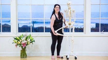
So as the song goes, the leg bone's connected to the hip bone, but I think we can go a little deeper. So we're going to be looking at the lower appendage. You have this gorgeous, longest bone of the body, this femur bone that we took a look at in an earlier chapter. Some points of reference on this bone, we have the femoral head, which snuggles into the socket of the pelvis or the acetabulum. Now this is a deeply set joint, so deeply congruent, almost like an orange sitting inside of a coffee cup.
A lot of the surface of the orange touches a lot of the coffee cup, so a lot of the femoral head touches a lot of the acetabulum, making a really strong, secure, snuggly fit. You also have a bunch of muscles and broad sheets of ligaments in this region, making it a very secure, stable joint. A ball and socket joint can take many different movements. It's one of the most freely movable joints. So it can do flexion, it can do extension, it can do abduction and adduction, it can do external rotation and internal rotation, and it can do kind of a combo, poo-poo platter of circumduction or circling because of that ball and socket joint.
We have the neck of the femur, and then we can find our greater trochanter, this outer bump bone. If you take your hands to the outer part of your upper thigh and just kind of feel around, and then just turn your leg, external rotation, internal rotation, and you'll feel this little bump roll underneath your hand, that's your greater trochanter. So sometimes when students come in, they go, I have hip pain, and they point to their hips or my hips are sore, and they kind of point to this region or the tush region. That's a little less concerning to me than when people say, I have hip pain, and they point to the groin area. That's where the socket, the head of the femur, sits in the socket of the acetabulum.
That's more indicative of something going on in the joint, and because the joint is avascular, it doesn't have a good blood supply, it doesn't heal very well. So that's a little bit more interesting or concerning to me as a yoga teacher. Moving down this long shaft of the femur, it terminates here at the little epicondyles, the little bump bones on the outside of the lower femur. You can feel those on your own body, taking your hands to the end of the femur and feeling those bump bones on either side. Here is our friend, the patella, or the kneecap.
Remember, it's a sesemoid bone, which means you're not born with it. It's a rock-like bone that's formed inside of the quadricep tendon, so like a rock in a stocking. The stocking is attached, but the rock is not. So if you take your hands to your own patella, and you extend your leg out in front, not locked, not bent, kind of in the midway point, you should be able to kind of manipulate your kneecap a little bit medial and a little bit lateral if you have a little wiggle. Some people are grossed out by that, don't do it if you're grossed out.
Look away for a second. Moving down, we have one long bone, this upper leg bone, and then we have two lower leg bones. We have the tibia, which is on the medial side, this is the bigger of the two, and then the fibula, which is the smaller, more lateral bone. Now this one's a little bit more challenging to feel. If you take your hands to the front of your shin, you're going to feel this little bump, it's the tibial tuberosity, it's the potato of your shin, the little bump, and this is where the patellar tendon, the quadricep tendon that encases the patella attaches to, and sometimes that can be kind of knobby on the floor, it might need some padding if you're standing on your knees.
You might run your hand down the front of the shin and feel the sharper edge of the tibia, or on the inside medial edge, you might feel that little bulge of the top part of the tibia. The fibula is more challenging to feel. The fibula is smaller with a lot of muscles on top, so where you can really feel the fibula, maybe up here by the knee, but definitely on the outer bump bone of your ankle, that's the end of the fibula, the inner bump bone of your ankle, the end of your tibia. The tibia and the fibula form almost like a wrench, like one of those crescent wrenches that sit on top of some of the foot bones or tarsals that will allow the action of flexion and extension in your ankle joint, but it's called something fancier down there. In the ankle, it's called pointer flexion and dorsiflexion.
The top part of the foot is like a dorsal fin, the dorsal side. The bottom side, you might have heard of plantar fasciitis. The bottom side of the foot is called the plantar side, so we have plantar flexion or pointing dorsiflexion or flexing. Moving down the road here, we can see we have all of these different rock-like bones forming the tarsals of the foot. The most impressive by far is that calcaneus bone or heel bone, which you can grab when you grab your heel, that's what you're grabbing.
It's also where that gastrocnemius tendon attaches to the calcaneus of the very famous tendon called the Achilles tendon. Each one of these tarsals has their own name. For our sake, we don't really need to name them here. We can call them as a group, the tarsals. This top one that articulates with the tibia and fibula called the talus, and that's what makes that kind of monkey wrench action.
Moving down past the tarsals, we have the metta tarsals, making up kind of the mid part of your foot, which you have five on each foot. Then down here, we have the phalanges. It's just more fun to say it that way. It's really phalanges, but if you say phalanges, it feels a little bit nicer. Those phalanges, they're just so teeny tiny, you have three in each toe and two in the big toe.
If you look at your own foot, just take a moment to marvel at that teeny tiny pinky toe and it has three phalanges in the toe. It looks like a little grape, but it's quite impressive. Now that we know all of the bones of the legs, now we can slap some muscles on and talk about their action.


You need to be a subscriber to post a comment.
Please Log In or Create an Account to start your free trial.